Research

How does molecular organisation control tissue-scale function?

and

How do we measure that?

Overview of research interests and techniques in the FMD Lab

Summary

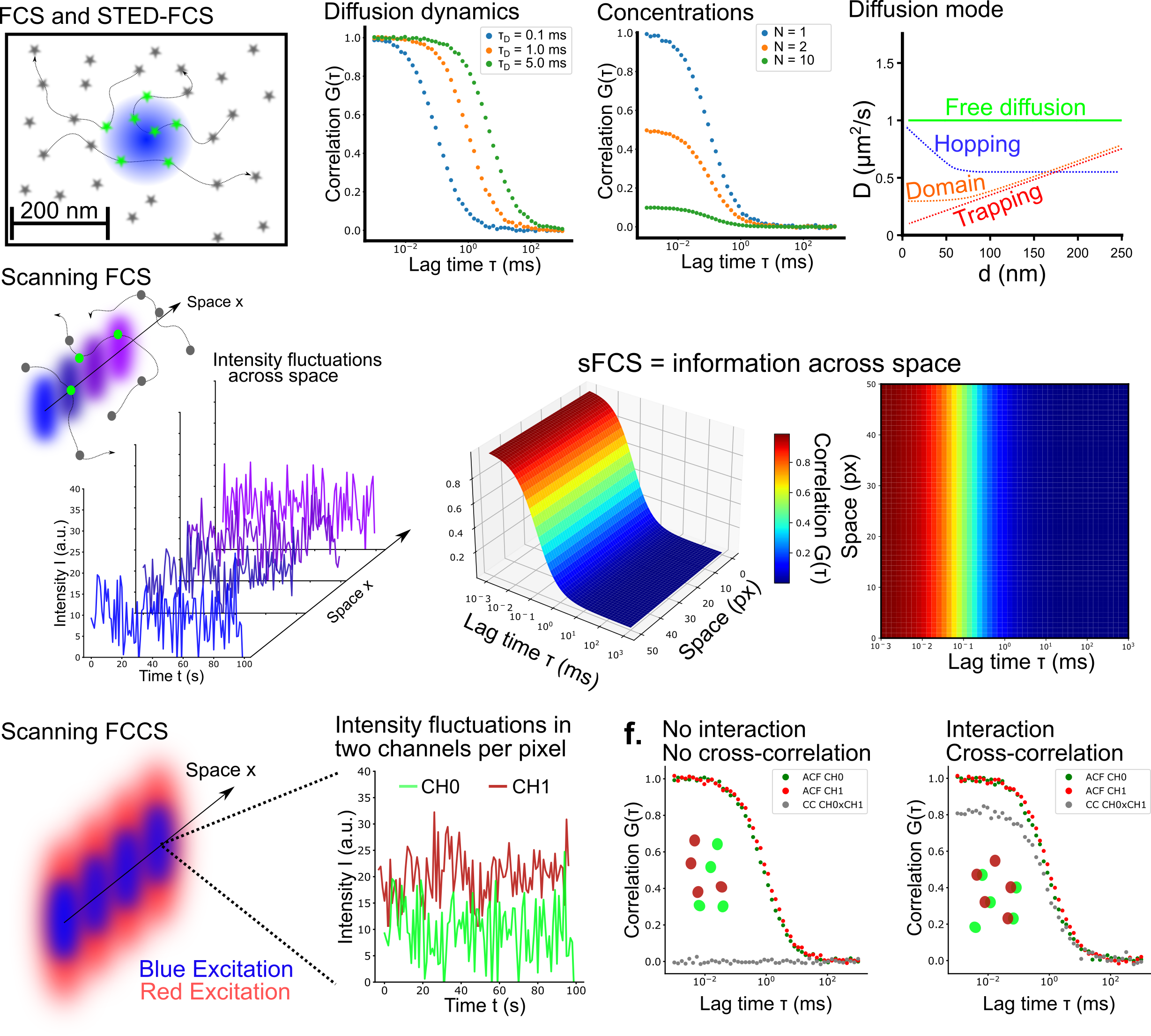
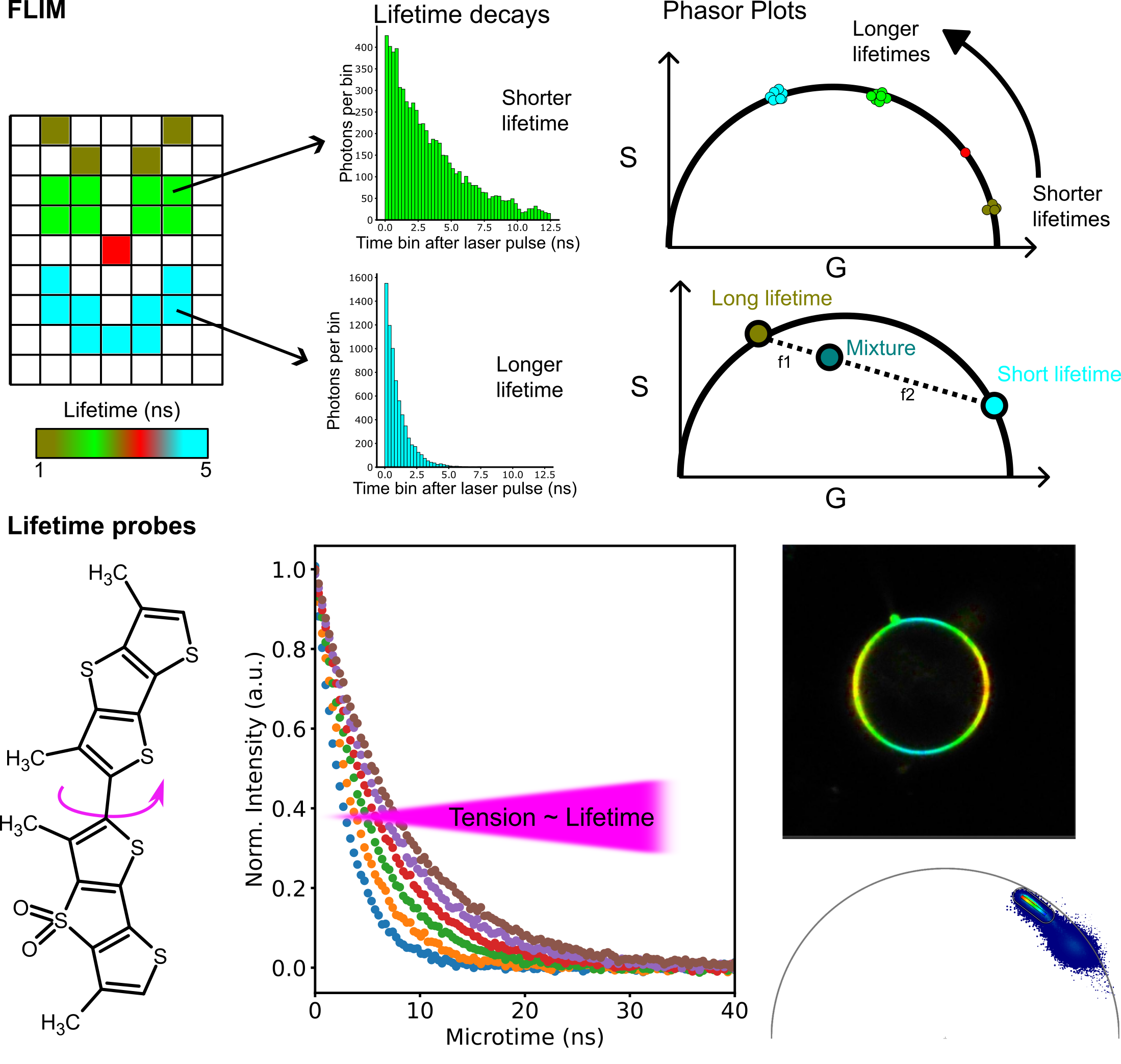
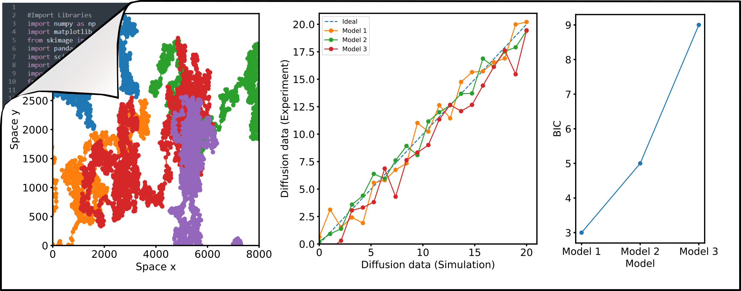
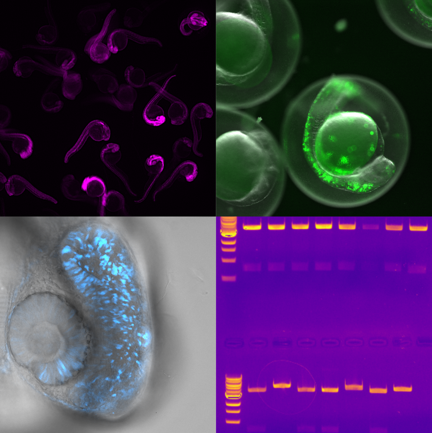
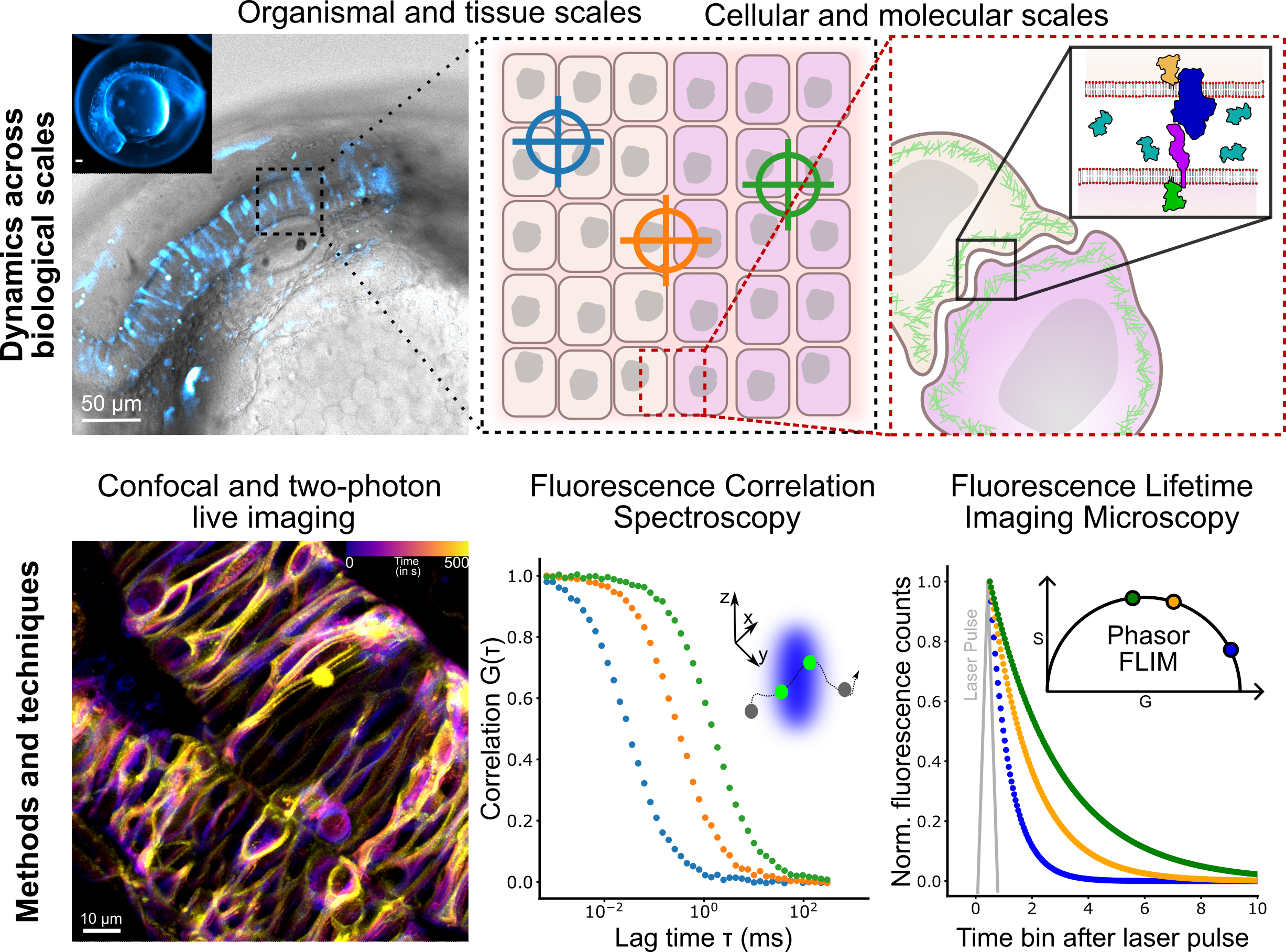
In the Fluorescence and Membrane Dynamics (FMD) Lab, we investigate how molecular motions and interactions give rise to complex organisation and function. Our research spans model systems of varying complexity. We work with artificial membranes with reconstituted proteins, cultured cells, and zebrafish embryos, enabling us to study dynamics across different biological scales. While in vitro systems provide a high degree of control, the physiological context of an organism offers unchallenged insights into the role of the native environment.

Our primary tools are based on advanced fluorescence microscopy. We develop and apply fluorescence correlation spectroscopy (FCS) and fluorescence lifetime imaging microscopy (FLIM) methods to make every photon count. Increasing sensitivity and specificity is especially important in deep tissue imaging. Currently, the lab focuses on studying the mechanisms underlying zebrafish hindbrain development, establishing methods to link nanometre-scale molecular dynamics, cellular signalling, and tissue-scale mechanics. Furthermore, we are developing a growing interst in probing metabolic changes using FLIM.

Key questions include:

1. How are lipid and receptor mobility in the plasma membrane influenced by tissue-scale mechanics?
2. How is the extracellular space organised, and how does it affect ligand delivery to membrane-bound receptors?
3. How are biophysical properties contributing to cellular identity within the tissue?

By bridging scales and developing cutting-edge fluorescence techniques and probes, the FMD Lab aims to uncover the fundamental biophysical organising principles that underpin complex biological systems and phenomena.

Technology & Resources

We use and develop a variety of tools.

Descriptive text

Fluorescence fluctuation spectroscopy techniques

Fluorescence fluctuation spectroscopy (FFS) and in particular fluorescence correlation spectroscopy (FCS) are powerful approaches to study molecular diffusion, concentration dynamics, and oligomerisation in living systems. Our research focuses on advancing and applying these techniques to probe molecular behaviour at the nanoscale and in complex systems. We have integrated FCS with stimulated emission depletion (STED) microscopy, creating STED-FCS, which enables direct measurements of diffusion dynamics with nanometre resolution. This combination allows us to map spatial variations in molecular mobility and identify local diffusion barriers, such as binding sites or membrane heterogeneities. While STED-FCS offers unmatched spatial resolution, it requires specialised instrumentation. To make nanoscale diffusion analysis more widely accessible, we are developing confocal scanning FCS (sFCS) pipelines that use advanced statistical analysis to extract hidden dynamic processes from ensemble measurements. By exploiting fluorescence cross-correlation spectroscopy (FCCS), we can study molecular interactions and co-diffusion. In combination with sFCS, this uniquely enables mapping of diffusion dynamics across space. Our current efforts focus on extending sFCS to reliably detect and quantify protein oligomerisation and clustering in living cells using single-colour measurements. In addition, FCS provides access to sub-microsecond time scales, allowing us to characterise the photophysical properties of fluorescent dyes in detail. In collaboration with probe developers, we use these insights to guide the design of improved fluorescent labels. Finally, we are developing robust and automated analysis pipelines to ensure consistent and reproducible FCS-based characterisation across diverse experimental conditions.

Key Publications:
Schneider et al., Nature Comms (2024)
Schneider et al., J Phys D Appl Phys (2020)
Sezgin et al., Nat Protoc (2019)
Schneider et al., Nano Lett (2018)
Schneider et al., ACS Nano (2018)

Descriptive text

Biophysical Imaging with Fluorescence lifetime imaging microscopy (FLIM)

Fluorescence lifetime imaging microscopy (FLIM) adds a new dimension to conventional fluorescence microscopy by measuring how long a fluorophore remains in its excited state after illumination. This time, known as the fluorescence lifetime, is independent of fluorescence intensity. In our lab, we use time-correlated single-photon counting (TCSPC) to record the precise delay between excitation and photon emission for each pixel. This enables us to visualise subtle molecular differences that intensity alone cannot reveal, and to distinguish or multiplex fluorophores with overlapping emission spectra. We apply the phasor approach to analyse FLIM data, providing an intuitive and model-free way to visualise lifetime distributions. This method helps us identify multiple fluorescent species, detect molecular interactions, and interpret complex environments directly from the data. FLIM also enables the use of environment-sensitive probes whose lifetimes change in response to factors such as pH, membrane tension, or ion concentration. By combining these “smart” sensors with advanced FLIM analysis, we gain quantitative insight into the biophysical properties and dynamics of living systems.

Descriptive text

Simulations and data analysis

Numerical simulations of diffusion dynamics, diffusion modes, and oligomerization are invaluable as ground-truth for the evaluation of new approaches. We aim to develop robust analysis and simulation pipelines utilizing open-source Python programming that can be adapted and used by anyone. We have contributed to the development and continue to support the FoCuS_scan software package which provides a graphical user interface to analyse sFCS data. In addition, We write scripts for image analysis in FIJI for fast, automated, and most importantly reproducible image analysis. Codes are available through Falk's Github page.

Key Publications:

Schneider et al., Nature Comms (2024)
Waithe et al., Methods (2018)
Schneider et al., ACS Nano (2018)

Descriptive text

Studying molecular interactions in physiological context

Fundamental biological processes, including signal transduction, cellular differentiation, and cell migration, hinge upon single molecule interactions within living systems. Yet, these interactions are often studied in isolated conditions, such as using reconstituted proteins in solution or transfected cells in culture. To delve into more intricate biological questions, it is essential to investigate these interactions within the full physiological context, specifically within tissues and whole organisms. Our recent research focuses on utilizing zebra fish as a model system to explore the influence of protein and nucleic acid interactions on early development. We employ standard and advanced molecular biology techniques, genetic modifications and we have initiated the optimisation of fluorescence fluctuation approaches and fluorescent probes for in vivo applications. This effort is geared towards enabling the observation of single molecule interactions using readily available instrumentation, facilitating the investigation of cellular signaling networks in developing tissues.

Key Publications:

Coming soon.